
|
| This cross section of the eyeball demonstrates a large white mass pushing into the vitreous. This is a retinoblastoma. This is what is seen as white on the fundusocopic examination. |
|
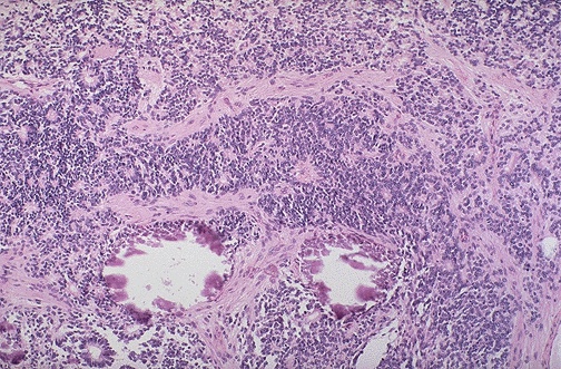
| Retinoblastoma is one of the "small blue cell tumors" of childhood. Necrosis and dystrophic calcification are commonly seen within this tumor. At low magnification, two small calcification can be seen below center. |
|
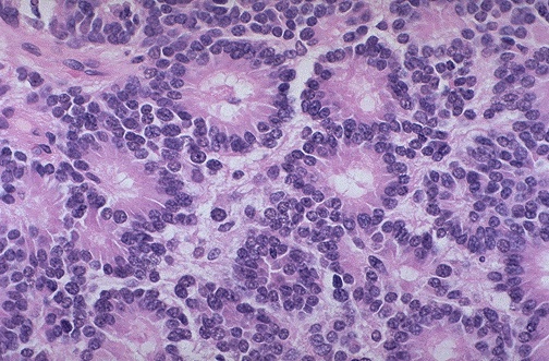
| Retinoblastoma is one of the "small blue cell tumors" of childhood. Necrosis and dystrophic calcification are commonly seen within this tumor. The characteristic microscopic pattern is arrangement of the small blue cells into Flexner-Wintersteiner "rosettes" as shown here. |
Examinations Photographs Movies Links Home Search noJava Home |